Mouse Csf1 ELISA KIT (96 T)
CNY 3,200.00
货期*
3周
规格
Specifications
| Product Data | |
| Description | Mouse M-CSF Immunoassay |
| Size | 96 T |
| Format | 96-well strip plate |
| Assay Type | Solid Phase Sandwich ELISA |
| Assay Length | 4 hours |
| Signal | Colorimetric |
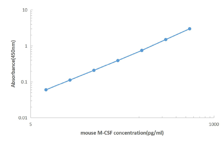
| Curve Range | 7.8-500 pg/ml |
| Sample Type | Serum, plasma, Cell culture supernatant |
| Sample Volume | 100 uL |
| Specificity | This assay recognizes both natural and recombinant mouse M-CSF. |
| Sensitivity | 4 pg/mL |
| Reactivity | Mouse |
| Interference | No significant interference observed with available related molecules. |
| Components |
|
| Background | M-CSF, also known as CSF-1, is a four-α-helical-bundle cytokine that is the primary regulator of macrophage survival, proliferation and differentiation. M-CSF is found as isoforms of various sizes. All isoforms contain the N-terminal 150 amino acid (aa) portion that is necessary and sufficient for interaction with the M-CSF receptor, but may vary in activity and half-life. Full length mouse M-CSF transcripts encode a 520 aa type I transmembrane (TM) protein that forms a 140 kDa covalent dimer. Differential processing produces two proteolytically cleaved, secreted dimers. One is an N- and O-glycosylated 86 kDa dimer, while the other is modified by both glycosylation and chondroitin-sulfate proteoglycan (PG) to generate a 200 kDa subunit. Human M-CSF is active in the mouse, but mouse M-CSF is reported to be species-specific. Sources of M-CSF include fibroblasts, activated macrophages, endometrial secretory epithelium, bone marrow stromal cells, vitamin D-stimulated osteoblasts, and activated endothelial cells. |
| Gene Symbol | Csf1 |
Documents
| Product Manuals |
Customer
Reviews
Loading...


 United States
 Germany
 Japan
Japan
 United Kingdom
 China
China

