Ki67 (MKI67) Rabbit Polyclonal Antibody
CAT#: TA336650
Rabbit Polyclonal Ki-67/MKI67 Antibody
Need it in bulk or conjugated?
Get a free quote
TA336650 is a possible alternative to TA336566.
CNY 6,056.00
货期*
5周
规格
经常一起买 (1)
beta Actin Mouse Monoclonal Antibody, Clone OTI1, Loading Control
CNY 300.00
CNY 1,430.00
Specifications
| Product Data | |
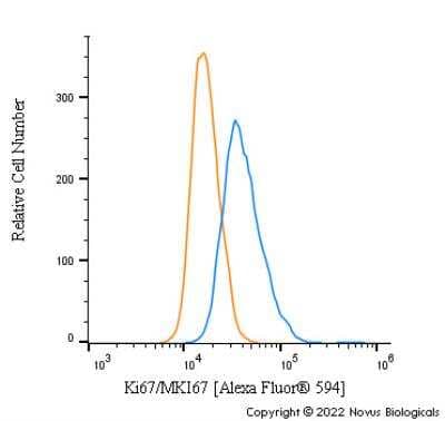
| Applications | FC, ICC/IF, IHC, Immunoblotting, IP, WB |
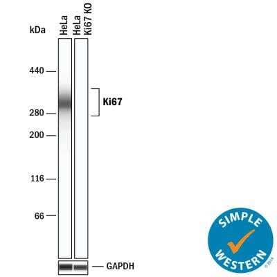
| Recommend Dilution | Flow Cytometry, Immunoblotting, Immunohistochemistry-Frozen: 1:50-1:300, Immunoprecipitation, Immunocytochemistry/ Immunofluorescence: 1:20-1:100, Western Blot: 1-2 ug/ml, Immunohistochemistry-Paraffin: 1:50-1:300, Knockout Validated, Immunohistochemistry: 1:50-1:300 |
| Reactivity | Human, Mouse, Rat, Porcine |
| Host | Rabbit |
| Clonality | Polyclonal |
| Immunogen | A synthetic peptide made to an internal region of human Ki67 (within residues 1550-1700) [Swiss-Prot# P46013] |
| Formulation | PBS, 0.05% Sodium Azide. Store at 4C short term. Aliquot and store at -20C long term. Avoid freeze-thaw cycles. |
| Concentration | lot specific |
| Purification | Immunogen affinity purified |
| Conjugation | Unconjugated |
| Storage Condition | Store at -20°C as received. |
| Gene Name | marker of proliferation Ki-67 |
| Database Link | |
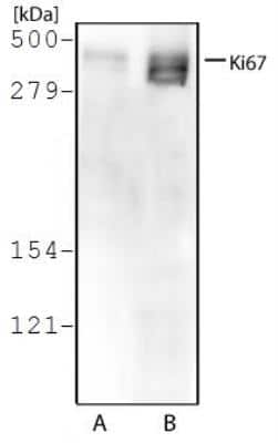
| Background | Originally discovered employing mouse monoclonal antibody against a nuclear antigen from Hodgkin's lymphoma-derived cell line, this non-histone protein was named Ki67 after researcher's location (Gerdes and colleagues), Ki for Kiel University in Germany and 67 referring to the clone number on the 96-well plate. It interacts with KIF15 as well as MKI67IP, and is suggested to be involved in cell cycle regulation. Ki67 is a large protein with expected molecular weight of about 395 kD and has a very complex localization pattern within the nucleus, one which changes during cell cycle progression. Its expression occurs specially during late G1, S, G2 and M phases of the cell cycle, while in cells undergoing G0 phase, Ki67 remains undetectable. Ki67 undergoes phosphorylation/dephosphorylation during mitosis, is susceptible to proteases and its structure implies that its expression is regulated by proteolytic pathways. Ki67 is associated with nucleolar DFC (dense fibrillary component) and its regulation appears to be tightly controlled (estimated half life is 60-90 min, regardless of the cell position in the cell cycle), presumably by precise synthesis and degradation systems involving proteasome, a protease complex. Due to its association with cell divison process, Ki-67 is routinely used as cellular proliferation marker of solid tumors as well as certain hematological malignancies, and a correlation has been demonstrated between Ki-67 index and the histopathological grade of cancers. |
| Synonyms | KIA; MIB-; MIB-1; PPP1R105 |
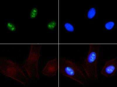
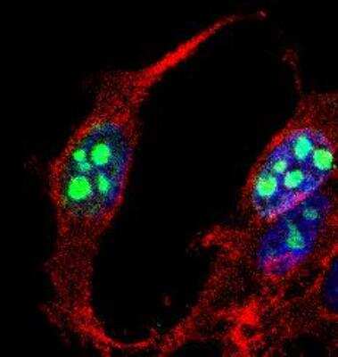
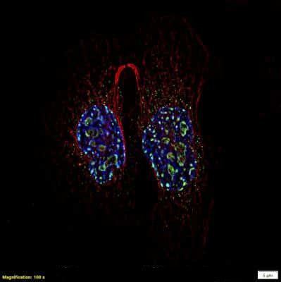
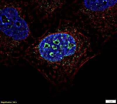
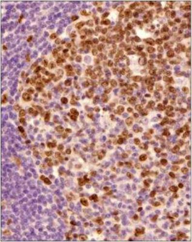
| Note | This Ki67 antibody is useful for Immunohistochemistry on frozen and paraffin-embedded sections and Immunocytochemistry/Immunofluorescence. There have been mixed results in Western blot; however, NB500-170 has been used successfully Western Blog reported by a customer review. *Formalin fixed paraffin embedded tissue sections require high temperature antigen unmasking with 10 mM citrate buffer, pH 6.0 prior to immunostaining. This antibody will not work without optimal antigen retrieval. This is probably the most critical step. NOTE: We suggest an incubation period of 30 minutes at room temperature and to use DAB to stain the protein (immunofluorescence may give problems as the protein is nuclear). |
| Reference Data | |
| Protein Families | Druggable Genome, ES Cell Differentiation/IPS |
Documents
| Product Manuals |
| FAQs |
| SDS |
Resources
| 抗体相关资料 |
Customer
Reviews
Loading...


 United States
 Germany
 Japan
Japan
 United Kingdom
 China
China
